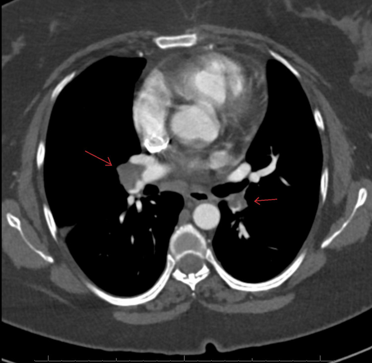
Figure 1. A CTA of the chest in axial view demonstrates acute bilateral pulmonary emboli involving the right pulmonary artery distally with extension into some of the right lower lobe and right middle lobe segmental and subsegmental arteries and some of the left lower lobe and left upper lobe segmental/subsegmental arteries and mildly enlarged pulmonary arteries.
CTA: computed tomography angiography

