Fig. 5.

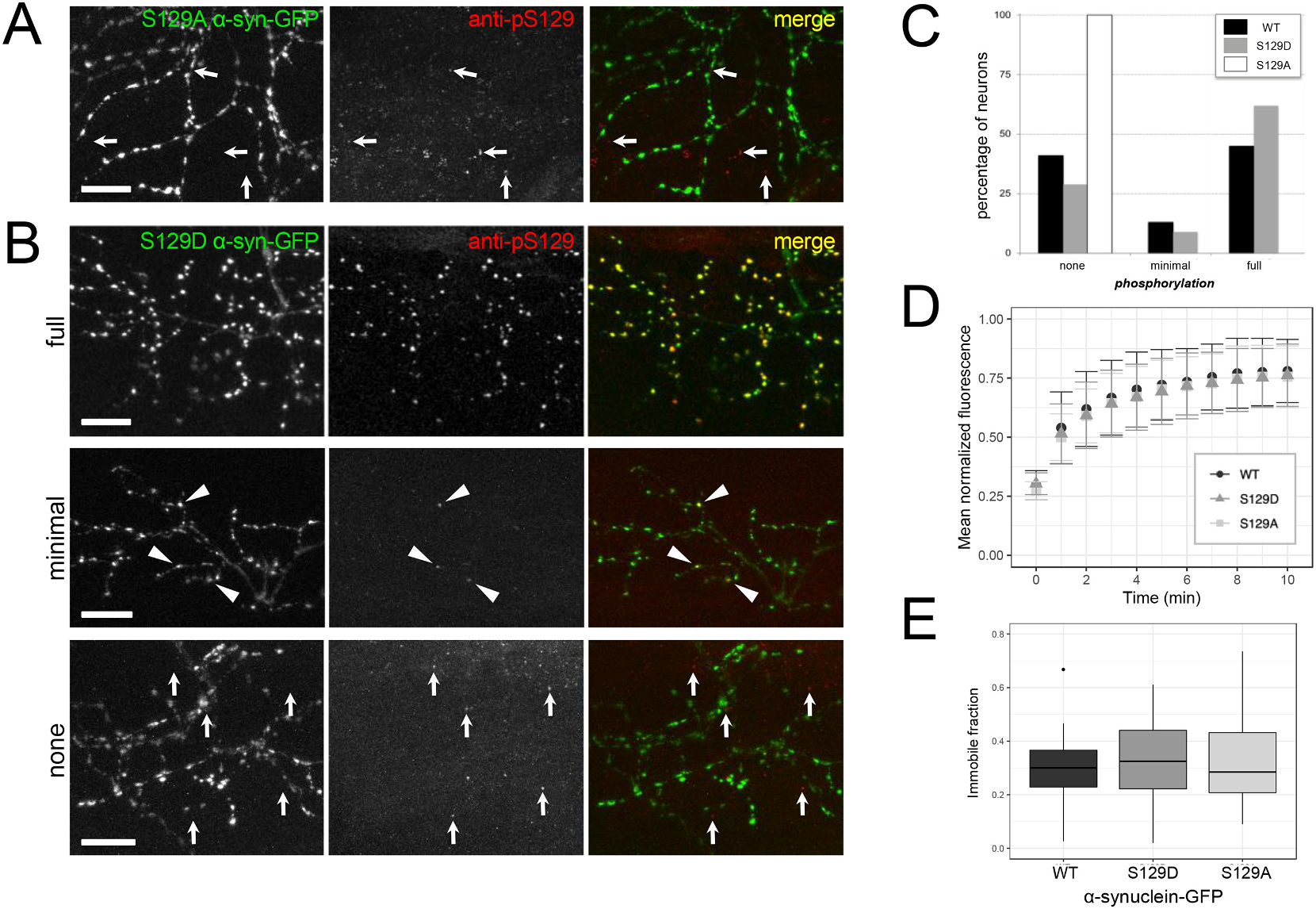
Genetic modification of serine 129 to alanine abolishes phosphorylation, but modification to alanine or aspartate does not affect immobile fraction.
A) S129A α-synuclein-GFP expressed in motor neurons and stained at 4 dpf does not exhibit phospho-S129 staining. GFP channel is shown at left, anti-phospho-S129 in the center, and merge at right. Arrows indicate immunostaining background puncta that are not overlapping with GFP channel. B) Similar to WT α-synuclein-GFP, S129D α-synuclein-GFP expressed in motor neurons and stained at 4 dpf is either fully phosphorylated throughout the axonal arbor (upper row example), minimally phosphorylated (middle row; a small fraction of terminals throughout arbor, indicated with arrowheads), or not phosphorylated (bottom row). In bottom row, arrows indicate immunostaining background puncta that are not overlapping with GFP channel. GFP channel is shown at left, anti-phospho-S129 in the center, and merge at right. Scale bars 10 μm; caudal is right and dorsal is up. All images are maximum intensity projections of stacks with the following depths: A) 39.2 μm B) 65.2 μm, 30.9 μm, and 66.6 μm (top, middle, and bottom respectively). C) Quantification of phosphorylation in motor neurons expressing each form of α-synuclein shows that neurons expressing wild type α-synuclein-GFP are distributed as 41.2% not phosphorylated, 13.2% minimally phosphorylated, and 45.6% fully phosphorylated (n = 69 neurons from 8 larvae). Neurons expressing S129D α-synuclein-GFP show a similar distribution with 28.6% not phosphorylated, 9.5% minimally, and 61.9% fully phosphorylated (n = 103 neurons from 14 larvae). In contrast, S129A α-synuclein-GFP expressed in motor neurons is not phosphorylated (100%; n = 130 neurons from 10 larvae). D) Group data of fluorescence recovery over time. Error bars represent standard deviation. Average tau for WT α-synuclein-GFP was 2.8 min (n = 26 terminals from 11 larvae); S129D was 2.7 min (n = 39 terminals from 13 larvae), and S129A was 2.3 min (n = 29 terminals from 7 larvae; one-way ANOVA, p = 0.638). E) The average immobile fraction for WT α-synuclein-GFP was 29.3% (n = 26 terminals from 11 larvae), S129D was 32.5% (n = 39 terminals from 13 larvae), and S129A was 32.8%; (n = 29 terminals from 7 larvae; one-way ANOVA, p = 0.641). In E, bars indicate range of data within 1.5 IQR; outlier outside this range shown as a point.
