
Fig. 4.
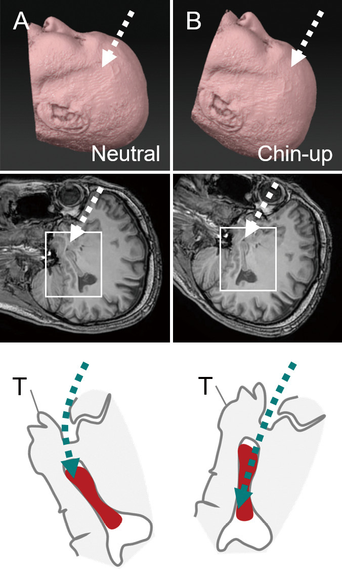
Chin-up position and surgical fields on the sagittal plain.
Schematic diagrams showing cases in the neutral (A) and chin-up (B) positions (upper row). The boxed area in the sagittal MR image, including the Sylvian fissure and hippocampus (middle row), is enlarged and schematized (bottom row). The hippocampus is drawn as a (red) solid object. The direction of the surgical trajectory is indicated by the (green) dashed lines. Note that the long axis of the hippocampus is on the straight surgical trajectory.