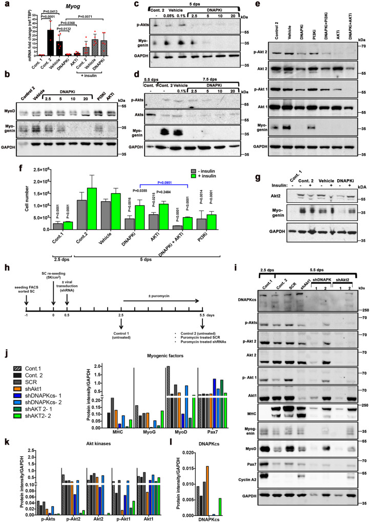
Fig. 2. DNA-PKcs is required for the expression of myogenin in a Akt-2 dependent manner.
a–g Myogenic cells treated with inhibitor of kinases. C2C7 cells were grown until 2.5 dps, following the scheme in Fig. S3d, and treated with inhibitors (alone or in combination, at the indicated doses) until 5 dps (panels a–c, e). SCs (d) were treated following the scheme in Fig. 1c, i.e. treated at 5.5 dps and collected at 7.5 dps. Control 1, untreated at 2.5 dps (or 5.5 dps, panel d); control 2, untreated at 5 dps (or 7.5 dps, panel d); vehicle, untreated (DMSO) at 5 dps (or 7.5 dps, d). a mRNA fold changes of Myog expression (n = 3–6) in C2C7 cells, mean ± SD normalized to the TBP-housekeeping gene, black columns, left part of the histogram. In the right part of the histogram, gray hatched columns indicate the same samples in the presence of 10 µg of insulin. Significance by ordinary one-way ANOVA (F = 9.943, DFn = 7, DFd = 30, p < 0.0001) with post-hoc Tukey’s multiple comparisons test, significant p values are indicated on the histogram. b Western blot (WB) of C2C7 cells treated with increasing doses of DNAPKi (NU7441), or 1 µM PI3Ki (ZSTK474), or 5 µM AKTi (MK2206); two bands of MyoD are present [72], and a band shift is observed at the highest doses of DNAPKi. c WB of Myogenin and p-Akts in C2C7 cells at increasing concentrations of DNAPKi. Two vehicle lanes (0.05% and 0.1%) correspond to the volume of DMSO used with 2.5 µM and 10 µM DNAPKi, respectively. d WB of Myogenin, Akts, and p-Akts of SCs in culture until 5.5 dps before treatment, with increasing concentrations of DNAPKi for two more days. e WB of C2C7 cells treated with 10 µM DNAPKi, PI3Ki (LY294002), and 5 µM AKTi, alone or in combination (left panels), and cultured in the presence or in the absence of inhibitor(s) until 5 dps; in panels b and e, vehicle contains 0.1% DMSO. In WBs, GAPDH was used as reference housekeeping protein. f C2C7 cell number and g WB of Myogenin and Akt2, upon growth and differentiation in the presence and in the absence of various inhibitors and insulin (green columns). As expected, insulin did not improve proliferation of cells treated with PI3Ki, because the PI3Ks are inhibited in this condition. In WB GAPDH was used as reference housekeeping protein. Significance by two-way ANOVA (F = 4.66, DFn = 18, DFd = 69, p < 0.0001), with post-hoc Tukey’s multiple comparisons of test vs. the corresponding vehicle conditions (vehicle or vehicle + insulin) when not otherwise indicated (P values shown on the histogram). h–l Analysis of myogenic factors and Akt kinases upon shRNA-dependent silencing of Akt1, Akt2, or DNA-PKcs. h Scheme and readouts of the experiment: SCs tested at 2.5 dps and 5.5 dps (control 1 and control 2, respectively) in the absence of puromycin (selection) treatment; SCR (scramble RNA) or shAkt1, shDNA-PKcs (2 independent clones), and shAkt2 (2 independent clones) at 5.5 dps upon puromycin selection. i WB of myogenic factors (differentiation markers: MHC (MF20) and Myogenin; myoblast marker MyoD, and stem cell marker Pax7), kinases (Akt1, Akt2, phospho-Akt1, phospho-Akt2, phospho-Akts, and DNA-PKcs), and the cell cycle marker cyclin A2. MyoD levels remained relatively high in SCR, perhaps as a consequence of the lentiviral and selection procedure. Normalization of protein levels with the reference protein GAPDH upon quantification of bands with Imagelab (BIORAD) for j myogenic factors, k individual Akt kinases and their phosphorylated forms, global p-Akts, and l DNA-PKcs. Uncropped gels are shown in “Original data file”.

