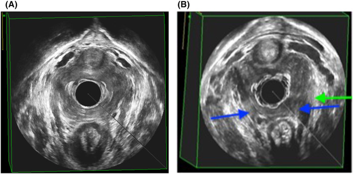
FIGURE 2.

(A) Example of an interpretable endovaginal ultrasound volume; levator ani muscle intact. (B) Example of an interpretable endovaginal ultrasound volume, LAD score 8p. Blue arrows indicating bilateral PP/PA defect (3 p each side) and green arrow indicating left‐sided PV‐defect (2 p).
