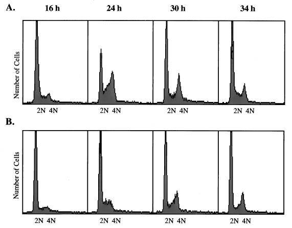
FIG. 1.
Cell cycle progression in confluent and subconfluent HDF cells. (A) Confluent plates of HDF cells were subcultured 1:6 and then placed in serum-free medium for 48 h. Medium containing 10% FBS was added to the arrested cells, which were harvested by trypsinization 16, 24, 30, and 34 h later. Cells were collected by centrifugation, fixed, and stained for flow cytometry as described in Materials and Methods. (B) Confluent plates of HDF cells were placed in serum-free medium for 48 h and then stimulated with medium containing 10% FBS. Cells were collected and stained at the same times as shown for panel A.

