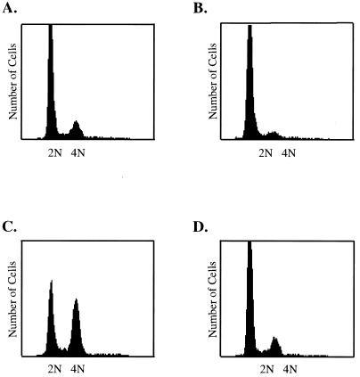
FIG. 6.
Induction of HDF cell cycle progression by small-t antigen in the presence of serum. Confluent monolayers of HDF cells were infected with Ad-ST or the small-t mutant virus Ad-C103S and then fed with fresh medium containing 10% FBS at the end of the infection period. Cells were harvested, fixed, and stained for fluorescence-activated cell sorter analysis at 32 h postinfection. The patterns shown are for serum-stimulated cells (A), cells infected with Ad-ST in the absence of serum (B), cells infected with Ad-ST in the presence of 10% serum (C), and cells infected with Ad-LT in the presence of 10% serum (D).

