FIG. 1.
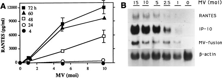
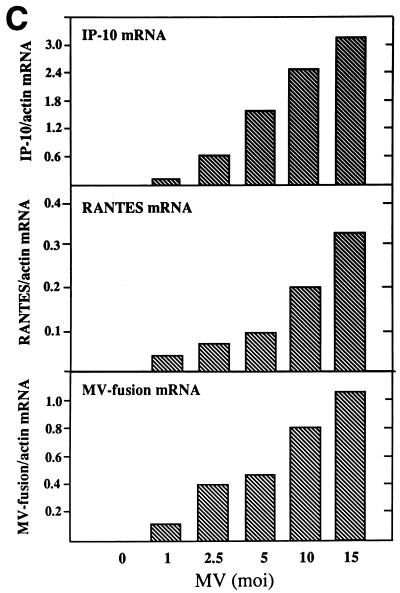
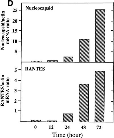
RANTES induction by MV in U373 cells. (A) RANTES protein. U373 cells (105/ml of 10% DMEM/well) in 24-well plates were infected with MV at an MOI of 1, 5, or 10 for 4, 24, 48, 60, or 72 h. Supernatants were then assayed for RANTES by ELISA. RANTES was not detected in mock-infected samples (MV at an MOI of 0). Data are means ± standard errors (SE) from three separate experiments performed in duplicate. (B) RANTES mRNA. U373 cells (107/75-cm2 flask) were infected with the indicated dose of MV for 48 h. Total cellular RNA was isolated and examined for RANTES mRNA by Northern blotting (20 μg of RNA/lane). MV fusion mRNA was used as an indicator of viral infection, and β-actin was used for normalization. The α-chemokine IP-10 was shown for comparison. Results represent one of two separate experiments. (C) The mRNA densities on an autoradiogram from panel B were quantitated, as described in Materials and Methods, and expressed as a ratio of the density of RANTES mRNA to that of β-actin mRNA. (D) Kinetics of RANTES mRNA expression. U373 cells were infected with MV at an MOI of 2.5, as in panel B, for the times noted. Total RNA was examined for RANTES and MV nucleocapsid mRNAs by Northern blotting (20 μg of RNA/lane). The mRNA bands on the autoradiogram were quantitated, and results are expressed as ratios of their densities to that of β-actin mRNA. A representative result from four separate experiments is shown.


