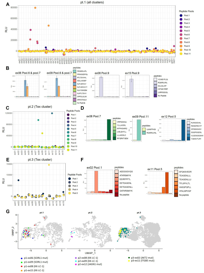
Figure 3.
Identification of tumor antigens recognized by CD8+ T cells. (A) The specificity of 70 TCRs from all 10 clusters for 41 predicted neoantigen peptides and 14 KK-LC-1 peptides tested by screening patient 1. All TCR clonotypes with more than one clone in each cluster were selected for testing. TCR TAP fragment/luciferase-transduced Jurkat (TCR TAP/luc-Jurkat) cells were co-cultured with autologous B-cell antigen presenting cells and pooled antigenic peptides (5–8 peptides/pool). Activation of TCR signaling was assessed by luciferase reporter assay driven by the NFAT-response element. (B) Individual peptides in positive pools 6, 7 and 9 were subsequently tested separately by further luciferase reporter assay. (C) In patient 2, the top 10 TCRs in descending order of frequency plus 5 TCR with one clone from the Tex cluster were tested against 14 predicted neoantigen peptides and 26 KK-LC-1 peptides. (D) The individual peptides in the positive pools were subsequently tested separately. (E) In patient 3, the top 15 TCRs from the Tex cluster were tested against 30 predicted neoantigen peptides. (F) The individual peptides in the positive pools were subsequently tested separately. (G) All identified TCR clones (n=140) with the nine tumor-antigen specific TCRs in three patients were projected onto UMAPs. UMAP, Uniform Manifold Approximation and Projection; TAP, Transcriptionally Active Polymerase Chain Reaction; TCR, T-cell receptor; Tex, exhausted T; RLU, relative light unit.

