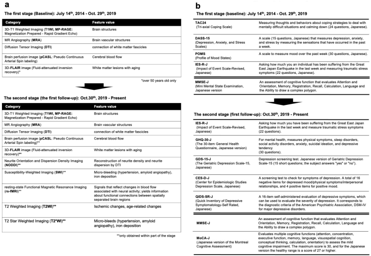
Figure 7.
MRI sequences and neuropsychological assessments during the first (baseline) and second (first follow-up) surveys.
a. The figure presents MRI sequences in the first (baseline) and second (first follow-up) surveys. The first survey (baseline) included 3D T1-weighted imaging (T1W1; MP-RAGE), magnetic resonance angiography (MRA), diffusion tensor imaging (DTI), brain perfusion imaging (pCASL), and 3D-FLAIR sequences. The second (first follow-up) survey has added neurite orientation and dispersion density imaging (NODDI), susceptibility-weighted imaging (SWI), resting-state functional magnetic resonance imaging (rs-fMRI), T2-weighted imaging (T2WI), and T2*-weighted imaging (T2*WI).
b. The figure presents questionnaires and on-site (interview) assessments in the first (baseline) and second (first follow-up) surveys at the top and bottom tables, respectively. Questionnaires are shown above the double line, whereas on-site assessments are shown below the line. In both surveys, the participants completed questionnaires assessing stress, psychiatric, personality, and neuropsychological factors at home and brought the completed forms with them on the day of the visit. On-site neuropsychological testing using the MMSE-J has been administered by trained testers. In the second (first follow-up) survey, the Montreal Cognitive Assessment, Japanese version (MoCA-J), is now administered to all participants.

