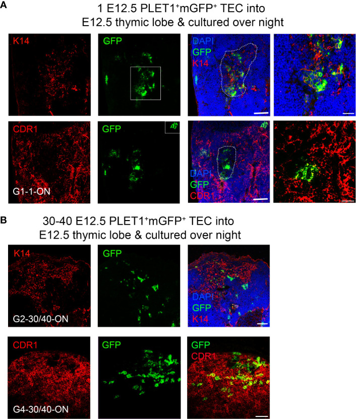
Figure 6.

A bipotent thymic epithelial progenitor cell is present after overnight culture. E12.5 mGFP+PLET1+ TEC were isolated from E12.5 thymi and a single cell was then injected into each wild type E12.5 thymus primordium; injected primordia were cultured overnight before grafting under the kidney capsule of recipient mice, grafts were recovered after 2-3 weeks and analysed with the markers shown. (A) Representative mGFP+ foci derived from a single cell showing co-localisation of mGFP+ cells with the mTEC marker Keratin 14 (k14) and the cTEC marker CDR1. DAPI shows nuclei. Right hand panels show higher magnification of boxed regions. (B) Representative mGFP+ foci from grafts of E12.5 thymic lobes injected with 40 mGFP+Plet1+ TEC and then cultured for 24 hours before grafting. Images show immunostaining for anti-K14 and CDR1 in two separate grafts. Graft name corresponds with graft identification in Table 3 . Scale bars (A) 150μm except right hand panels, 55μm, (B) 150μm. N for each condition represents an independent graft and is as indicated in Table 3 ; at least three biologically independent replicates were performed for each injection condition.
