FIG. 6.
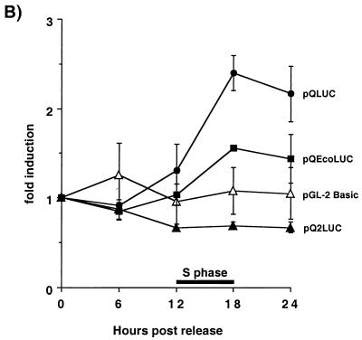
Qp activity is induced during S phase and is dependent on the E2F sites within the Q locus. (A) Structure of the Qp luciferase (LUC) reporter constructs. The bent arrow marks the Qp start site; the open square is the ISRE; black ovals and hatched rectangles are the EBNA-1 and E2F binding sites, respectively, within the Q locus. The upstream and downstream E2F sites are designated QpE2Fa and QpE2Fb, respectively, in Fig. 1 and the text. The nuleotide sequence of the QpE2Fb site (boxed sequence) in pQLUC and pQEcoLUC is shown at the right of each reporter construct. The three mutated base pairs within pQEcoLUC are underlined. (B) Fold induction of the Qp luciferase reporter constructs and the vector control, pGL-2 Basic. β-Gal assays were performed on each time point to normalize the amount of protein lysate used in the luciferase assays. The time of S phase is shown as a black bar spanning from 12 to 18 h after release from serum starvation.


