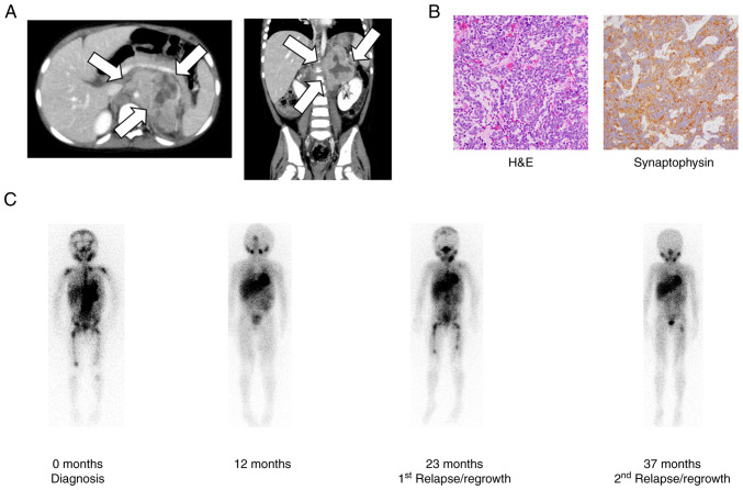
Figure 1.
(A) CT images of an abdominal tumor at diagnosis. A tumor with a diameter of ~6 cm (white arrows) was detected in the left adrenal gland. (B) Pathological examination of the adrenal tumor. Small round tumor cells were arranged in nests separated by slender fibers (original magnification, ×200). Immunostaining was positive for synaptophysin (original magnification, ×400). (C) Representative MIBG images during the entire course of treatment. MIBG-avid lesions were detected in left adrenal gland, right and left upper carpal bones, spine, pelvis, and right and left thigh bones at 0 months, disappeared at 12 months, and reappeared in the left upper carpal bones, spine, pelvis, and right and left thigh bones at 23 months and in the left thigh bone at 37 months. H&E, hematoxylin and eosin staining; MIBG, metaiodobenzylguanidine.

