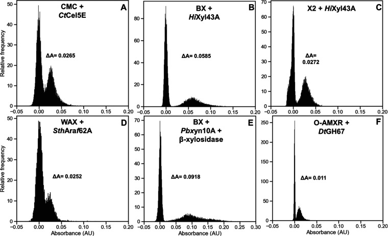
Figure 5.
Detection of CAZyme-expressing cells in droplets using the coupled assays. Droplets containing E. coli BL21 (DE3) cells transformed with plasmids encoding CtCel5E (A), HiXyl43A (B, C), SthAraf62A (D), PbXyn10A (E), or DtGH67 genes (F) were generated using the flow-focussing device shown in Supporting Figure S4C. After cell growth and protein expression, these droplets were picoinjected with CMC + glucose cascade, beechwood xylan + xylose cascade, xylobiose + xylose cascade, wheat arabinoxylan + arabinose cascade, beechwood xylan + β-xylosidase + xylose cascade or uronic acids + glucuronic acid cascade, respectively. Droplets were incubated at 25 °C for 48 h (A), 37 °C for 2 h (B, C, D, F), or 37 °C for 24 h (E), prior to absorbance measurement with the sorter shown in Supporting Figure S4E.

