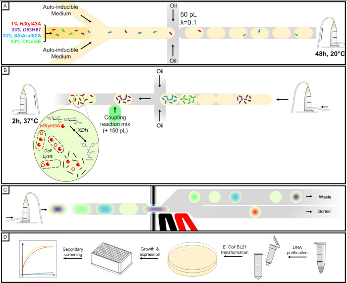
Figure 6.
Functional screening workflow of xylosidase activity in mixtures of droplets expressing two different CAZymes. (A) Cells expressing HiXyl43A, DtGH67, SthAraf62A, and CtCel5E were mixed to a 1:33:33:33 ratio and encapsulated into 50 pL droplets with λ = 0.1. (B) After a 48 h incubation allowing cell growth and protein expression, the droplets are picoinjected with a 150 pL coupling reaction mix allowing cell lysis and detection of xylose activity. (C) After a 2 h incubation allowing signal development, the droplets are sorted by AADS. (D) Positive droplets are collected, and the DNA they contain is extracted and purified, cloned into E. coli BL21 cells, and grown on a selective medium. 96 individual colonies are grown in autoinducible medium allowing protein expression. After centrifugation of the induced cells, the supernatant is assayed for xylose release from xylobiose in a microtiter plate.

