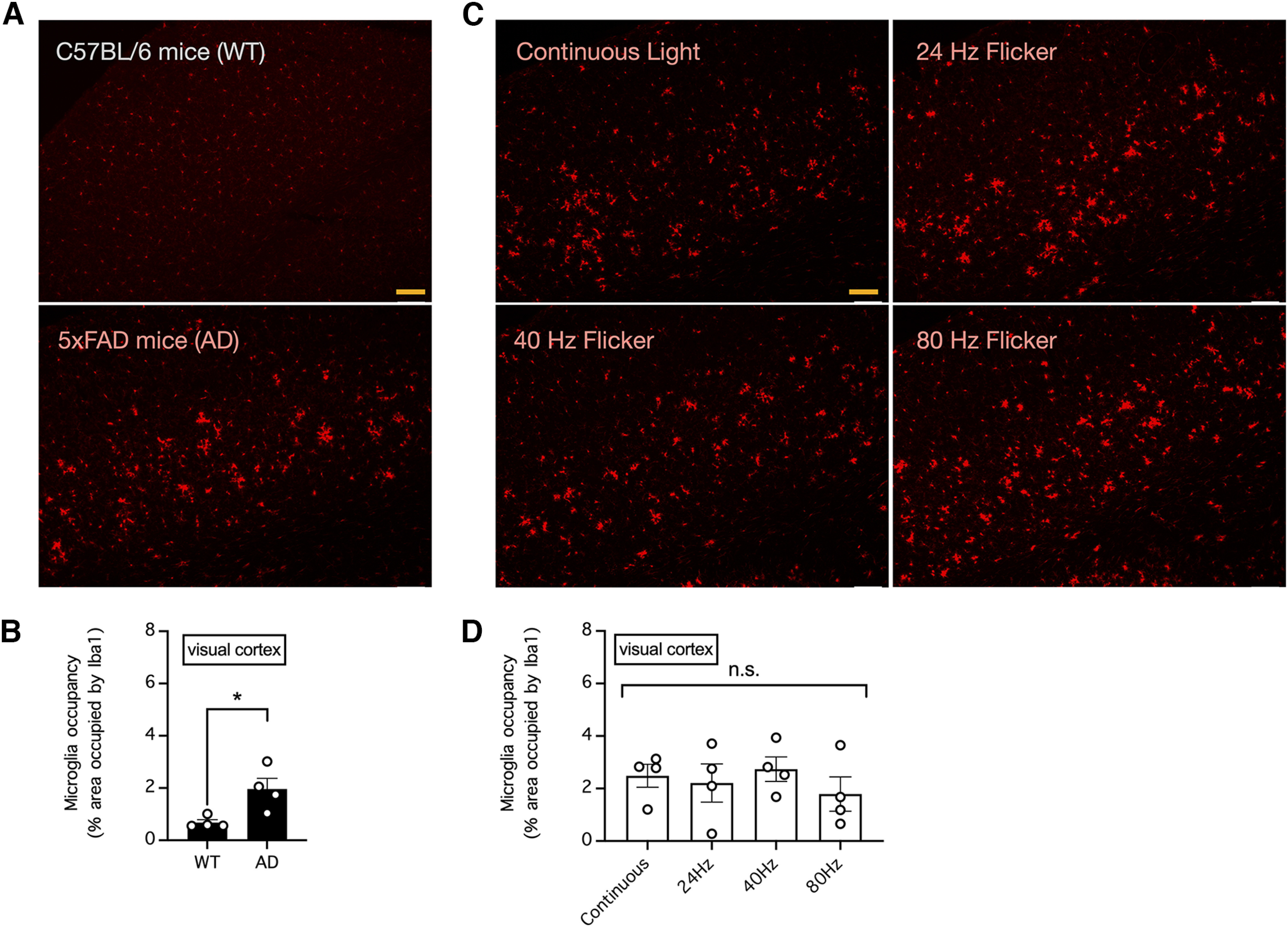
Figure 1.

Visual stimulation with light flickering at 40 Hz had no effect the area occupied by microglia in the mouse visual cortex. A, The relative area occupied by microglia in the visual cortex of 33-week-old control C57BL/6 and 5xFAD mice was determined by Iba1 immunostaining. Bar indicates 200 μm. B, Summary of the results in A showing the relative area occupied by microglia in the visual cortex. n = 4 mice per group. * indicates p < 0.05 when compared by unpaired t test. C, The relative area occupied by microglia in the visual cortex of 5xFAD mice subjected to daily 1-h visual stimulation with continuous light or light flickering at 24, 40, or 80 Hz for five weeks was determined by Iba1 immunostaining. Bar indicates 200 μm. D, Summary of the results in C showing the relative area occupied by microglia in the visual cortex. n = 4 mice per group. n.s. indicates no significant difference when compared by one-way ANOVA.
