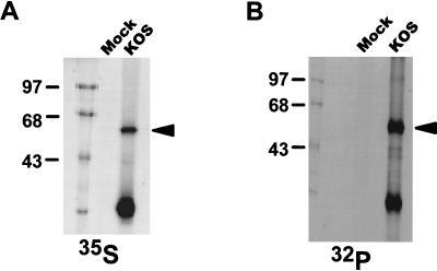
FIG. 1.
Immunoprecipitation of [35S]methionine- or 32Pi-labeled ICP27 from nuclear extracts of HSV-1-infected cells. RSF cells were either mock infected or infected with wild-type HSV-1 strain KOS. Cells were labeled with [35S]methionine (A) or 32Pi (B) in vivo for 5 h, after which the cells were harvested and nuclear extracts were prepared. The extracts were immunoprecipitated with anti-ICP27 monoclonal antibodies H1113 and H1119. The antigen-antibody complexes were separated on an SDS-polyacrylamide gel and detected by autoradiography. The bands corresponding to full-length ICP27 are indicated by arrowheads. The positions of protein molecular weight markers are shown on the left (in kilodaltons).

