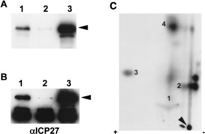
FIG. 4.
Two-dimensional tryptic phosphopeptide mapping of ICP27 from transfected cells. RSF cells were either infected with KOS or transfected with plasmid pCMV-ICP27 expressing ICP27 and labeled with 32Pi for 5 h. (A and B) Immunoprecipitated ICP27 from either a nuclear extract of infected cells (lanes 1), a cytoplasmic extract of infected cells (lanes 2), or a nuclear extract of transfected cells (lanes 3) was resolved on an SDS-polyacrylamide gel and transferred to a nitrocellulose membrane. The position of ICP27 is indicated by arrowheads. (A) The radioactively labeled proteins on the blot were detected by autoradiography. (B) The blot was subsequently probed with anti-ICP27 monoclonal antibodies (αICP27). The dark band seen in all lanes at 55 kDa is heavy-chain immunoglobulin G which reacts with the secondary antibody used in Western analysis and is present because immunoprecipitated proteins were transferred to the membrane. (C) Immunoprecipitated ICP27 from a nuclear extract of transfected cells (lanes 3, panels A and B) was subjected to two-dimensional tryptic phosphopeptide mapping as described in the legend to Fig. 3. The arrowhead indicates the sample origin on the TLC plate. The four major phosphopeptides are identified by numbers.

