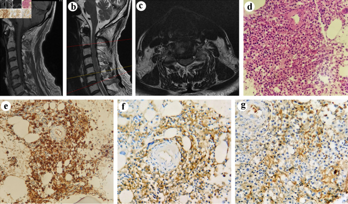
Figure 1.
MRI and pathological images of a 71-year-old male with MM after cervical spine surgery. Tl-weighted scan (a) and T2-weighted gradient echo scan (b) of cervical spine showed the morphology of the bone and spinal cord. Axial T2-weighted MRI of cervical spine showed C5/6 intervertebral disc protrusion (c). (d) The morphology of the tumor cells (H&E × 400). (e-g) Immunohistochemical findings: (e) the tumor cells expressing CD38 (original magnification × 400) and (f) kappa (original magnification × 400), with (g) lambda (original magnification × 400) rarely expressed. MRI: magnetic resonance imaging; MM: multiple myeloma; H&E: hematoxylin and eosin stain.

