Figure 1.

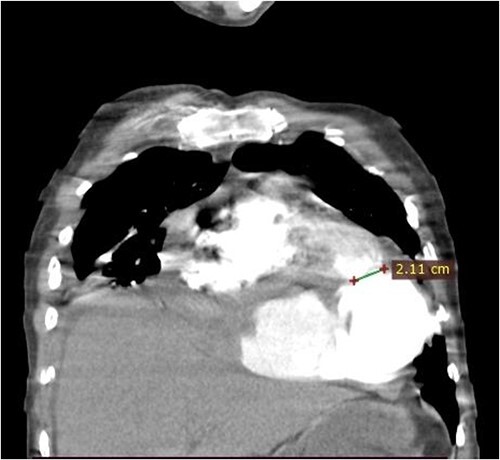
Coronal CT scan image. The figure shows a 2.1 cm communication between left ventricle and pseudoaneurysm. The heart is pushed upwards.

Coronal CT scan image. The figure shows a 2.1 cm communication between left ventricle and pseudoaneurysm. The heart is pushed upwards.