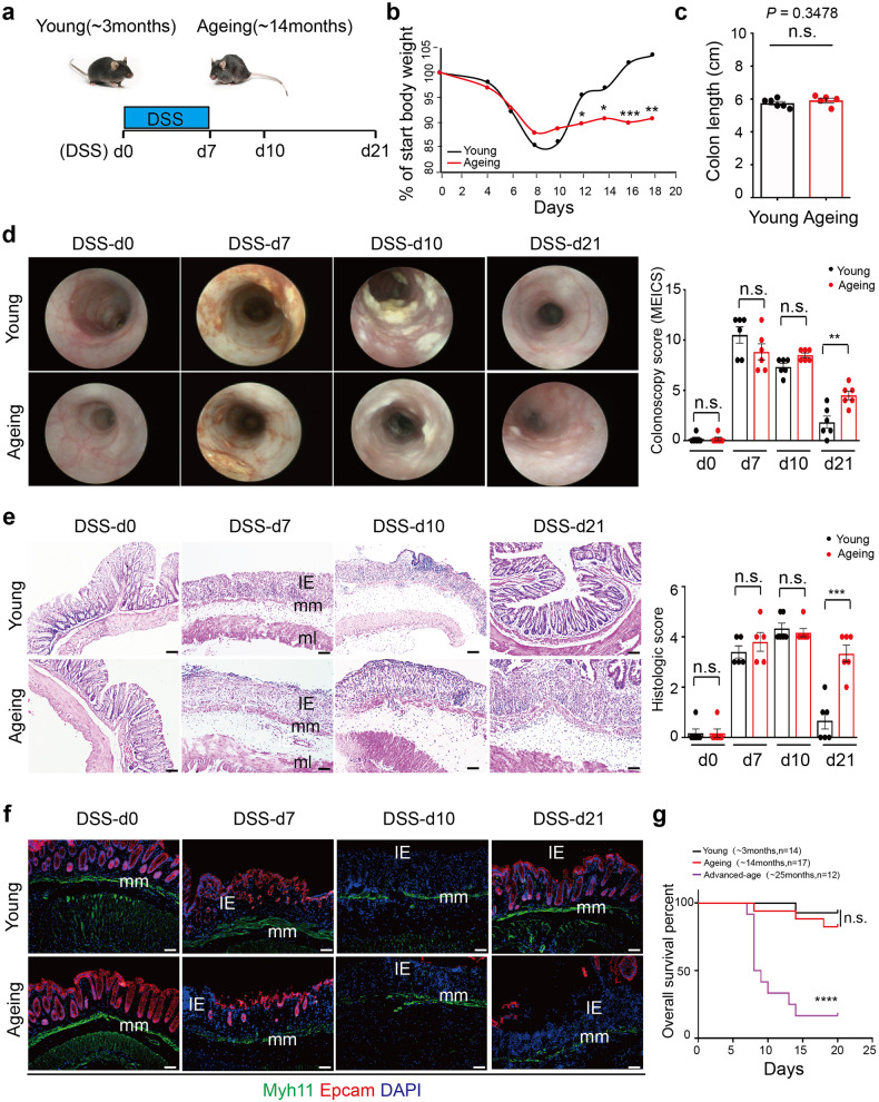
Fig. 1.
Decreased intestinal wound healing with ageing in DSS-induced acute colitis. a Schematic overview of DSS-induced acute colitis. Colon tissues were collected at the early phase of colitis at DSS-d7, during colitis at DSS-d10, and after the wound healing at DSS-d21. b The weight change in young and ageing mice during DSS-induced acute colitis. Young, n = 12; Ageing, n = 9. c The colon length of young (n = 6) and ageing (n = 5) mice at DSS-d7, mean ± SD. d High-resolution endoscopic images of young and ageing colon at 0, 7, 10 and 21 days after the beginning of DSS feeding. The severity of colitis was measured using the coloscopy score system (MEICS). n = 6 for each group, mean ± SD. e Representative H&E staining of colon tissues at 0, 7, 10 and 21 days after the beginning of DSS feeding. IE intestinal mucosa epithelium; mm muscularis mucosa; ml muscular layer; these abbreviations apply to the whole study. The histopathological grading of inflammation in young and ageing mice was recorded. DSS-d0, n = 6; DSS-d7, n = 5; DSS-d10, n = 6; DSS-d21, n = 6. Data are mean ± SD. Scale bars, 50 μm. f Immunofluorescence images of the temporal re-epithelialization process that occurs following the DSS-induced colitis in young and ageing mice. Sections were immunolabeled with Epcam (marker of epithelium cells) and Myh11 (marker of smooth muscle cells). Scale bars, 50 μm. g Survival analysis of the advanced-age (~25 months) mice (purple solid line; n = 12) compared to that of young mice (black solid line; n = 14; P < 0.0001 by log-rank test) during DSS-induced acute colitis. Note that survival of young and ageing (~14 months, red dashed line, n = 17) mice had no significant difference

