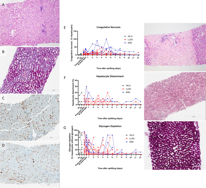
Fig. 3. Histopathology analysis of liver core biopsies taken throughout long-term perfusion of human split livers.
Slides were stained with haematoxylin and eosin to assess architectural integrity (A), Periodic acid-Schiff for glycogen depletion (B) and ki67 for cellular proliferation (C, D). The amount of cellular proliferation decreased from early in perfusion (C) to later in perfusion (D). Assessment of each slide was performed by a blinded specialist pathologist for coagulative necrosis (E), hepatocyte detachment (F) and glycogen depletion (G)22. Levels of coagulative necrosis and hepatocyte detachment remained low until the point of organ failure (E, F). Glycogen deposition within hepatocytes increased with long-term perfusion (G).

