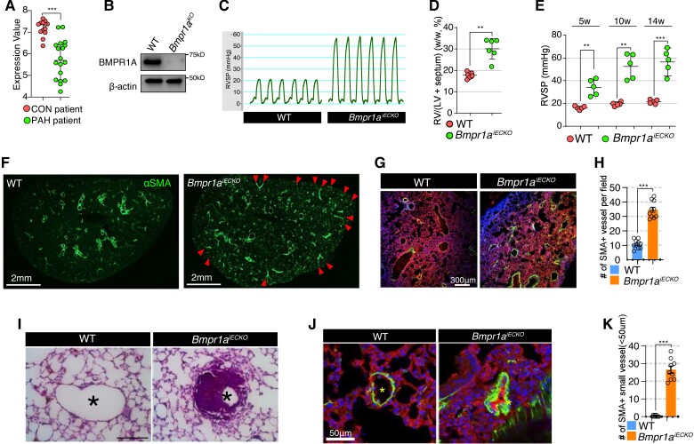
Figure 1.
Loss of endothelial BMPR1A leads to pulmonary arterial hypertension. (A) Microarray-based gene expression profile of BMPR1A in human lungs from control and PAH subjects. ***P < 0.0001 (unpaired t-test). (B) Western blot showing BMPR1A expression level in lung ECs from Bmpr1aiECKO mice with (Bmpr1aKO) or without (WT) Cre activation. (C) Right ventricular systolic pressure in Bmpr1aiECKO mice in comparison with control mice (n = 6). (D) The weight of right ventricle is significantly increased in Bmpr1aiECKO mice (dots) compared with wildtype mice (dots). **P ≤ 0.005 (unpaired t-test). (E) The right ventricular systolic pressure is progressively elevated over 14 weeks in Bmpr1aiECKO mice (dots) after tamoxifen administration compared with wildtype mice (dots). [*** < 0.0001, **P ≤ 0.005 (unpaired t-test), n = 5]. (F) Immunostaining showing overall αSMA alteration with vibratome section (150 μm thickness). αSMA deposition was significantly elevated in Bmpr1aiECKO mice (left). Arrowheads indicate αSMA-positive pulmonary perivasculature (scale bar = 2 mm). (G) Confocal images showing CD31 and αSMA Immunostaining in the lung of control (left) and Bmpr1aiECKO (right) mice (CD31; αSMA; DAPI). Note that αSMA-positive vessels were increased in Bmpr1aiECKO mice compared with wildtype mice (scale bar = 300 μm). (H) Quantification of αSMA-positive arteries per field in the lungs of control and Bmpr1aiECKO mice [***P < 0.0001 (unpaired t-test), n = 10]. (I) Haematoxylin and eosin staining showing medial hypertrophy of pulmonary artery in Bmpr1aiECKO (right) mice (scale bar = 100 μm). (J) Confocal images showing the muscularized and occluded small (<50 μM) arteries (αSMA; CD31; DAPI) in Bmpr1aiECKO mice (scale bar = 50 μm). (K) Quantification of small αSMA-positive arteries (<50 μM) in the lungs of control and Bmpr1aiECKO mice [***P < 0.0001 (unpaired t-test), n = 9].

