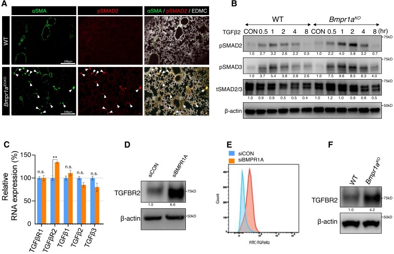
Figure 3.
Increased TGFBR2 expression in the absence of endothelial BMPR1A. (A) Immunostaining showing Increased pSMAD2 deposition in lung tissues from Bmpr1aiECKO (bottom) mice compared with control (top). White arrows indicate pSMAD2 and αSMA double-positive cells. (scale bar = 500 μm). (B) Western blot showing that mouse lung endothelial cells with Bmpr1a deletion were more sensitive to TGFβ2 stimulation compared with those from control cells. (C) RNA expression of Tgfbr2 was significantly increased while the expression of other TGFβ signalling components was either slightly decreased or unaltered in BMPR1A siRNA-treated PAECs compared with control siRNA-treated PAECs. [**P ≤ 0.005 (unpaired t-test), n = 3]. (D) Western blot showing that TGFBR2 expression was increased in BMPR1A siRNA-treated PAECs compared with control siRNA-treated PAECs. (E) Flow cytometry showing a significant increase of TGFBR2 expression in BMPR1A siRNA-treated PAECs compared with control siRNA-treated PAECs. (F) Western blot showing increased TGFBR2 expression in mouse lung endothelial cells with Bmpr1a deletion compared with those from control cells.

