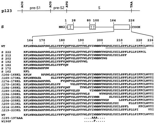
FIG. 1.
Schematic representations of S protein mutants. Plasmid p123 and S protein are depicted by horizontal thin lines. The HBV envelope protein open reading frame is divided into pre-S1, pre-S2, and S domains. Single base substitutions converted ATG (Met) start codons to ACG (Thr) codons for expression of the S protein only. Open boxes represent hydrophobic transmembrane regions in the S protein. Transmembrane signals I and II are indicated. The name and sequence of each mutant, including the positions of the first and last residues of the deleted sequence, are indicated. The letters KL correspond to an insertion of a Lys-Leu sequence at the site of deletion. The lower rows correspond to triple and single amino acid mutants. The position and nature of the substitutions are indicated. Underlined sequences represent putative transmembrane alpha-helices.

