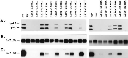
FIG. 5.
Detection of HDV RNA-containing particles in culture fluids from cells cotransfected with pSVLD3 HDV plasmid DNA and WT or S protein mutant DNA. (A) Immunoblot analysis of S proteins extracted from culture fluids from 0.5 × 10⁶ HuH-7 cells at days 6, 9, 12, and 15 after transfection with a mixture of 0.6 μg of pSVLD3 plasmid DNA and 1.4 μg of WT, env-negative (EN), or mutant HBV plasmid DNA. Particles sedimented from 200 μl of culture medium were disrupted in Laemmli sample buffer containing 2% SDS and 2% β-mercaptoethanol. Proteins were separated on a 12% acrylamide gel, transferred to a polyvinylidene difluoride membrane and probed with anti-S antibodies (1:1,000). (B) Cellular RNA was extracted from HuH-7 cells harvested at day 15 posttransfection. Five micrograms of total RNA was separated on an agarose gel and analyzed for the presence of HDV RNA after transfer to a nylon membrane and hybridization to a genomic strand-specific 32P-labeled HDV RNA probe. Following hybridization, filters were washed, dried, and autoradiographed at −70°C for 16 h with an intensifying screen. The size expressed in kilobases of HDV genomic RNA is indicated. (C) Particles sedimented by precipitation in the presence of 9% PEG from 500 μl of culture medium were disrupted in RNAble, and RNA was purified. RNA was analyzed by agarose gel electrophoresis followed by transfer at nylon membrane and hybridization to a genomic strand-specific 32P-labeled HDV RNA probe as described for panel B.

