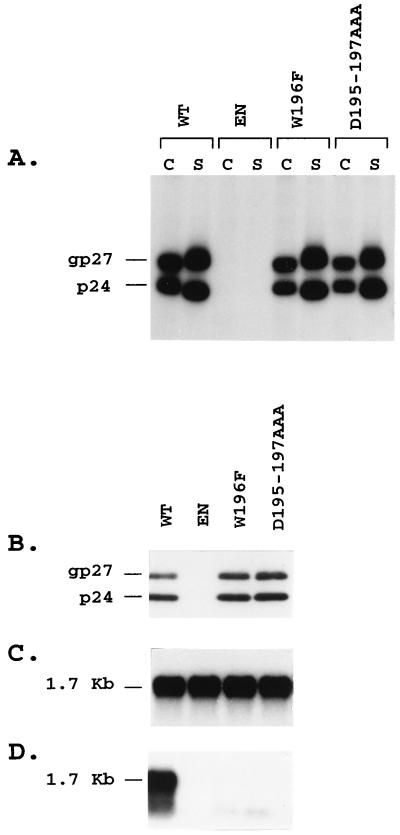
FIG. 6.
Analysis of WT and W196F and Δ195-197AAA S protein mutants for subviral and HDV particle secretion. (A) HuH-7 cells transfected with WT or S protein mutants were labeled with 200 μCi of [35S]Cys-[35S]Met for 24 h, and cell lysates (C) and supernatants (S) were immunoprecipitated with rabbit R247 anti-S antibodies. (B) For expression of HDV RNA-containing particles, 0.5 × 10⁶ HuH-7 cells were cotransfected with a mixture of 0.6 μg of pSVLD3 plasmid DNA and 1.4 μg of WT, env-negative (EN), or mutant DNA. Particles were harvested at days 6, 9, 12, and 15 posttransfection and sedimented by precipitation in the presence of 9% PEG from 200 μl of culture medium. Proteins were analyzed by SDS-PAGE and immunoblotting with anti-S antibodies (1:2,000). (C) Cellular RNA was extracted from HuH-7 cells harvested at day 15 posttransfection, and 5 μg was analyzed by gel electrophoresis and hybridization to a genomic strand-specific 32P-labeled HDV RNA probe. (D) RNA purified from particles precipitated from 500 μl of culture medium was analyzed by gel electrophoresis and hybridization to a genomic strand-specific 32P-labeled HDV RNA probe. The gel migration positions of intracellular or extracellular genomic HDV RNA (1.7 Kb) are indicated in panels C and D, and those of S proteins (p24 and gp27) are indicated in panels A and B. EN, env-negative.

