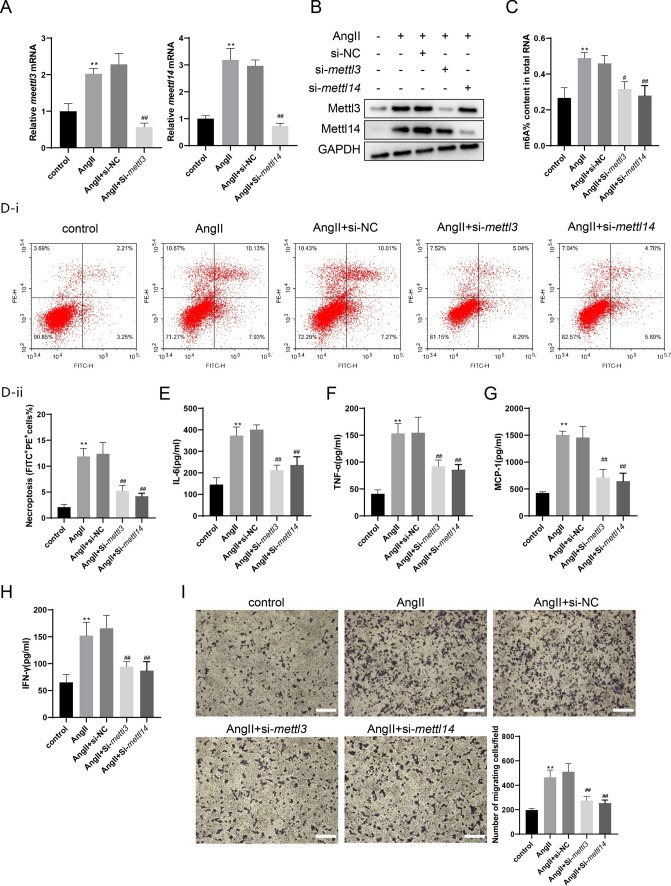
Fig. 2.
The interference with METTL3 or METTL14 reduced necroptosis and inflammatory response of Ang II-treated VSMCs. VSMCs were divided into the Control group and the Ang II group. In the Ang II group, VSMCs were incubated with 200 nM Ang II. VSMCs were also transfected with si-mettl3, si-mettl14, or NC (10 nM) for 24 h, followed by incubation with Ang II for 72 h. A The mRNA expressions of mettl3 and mettl14 were detected using the qRT-PCR. gapdh was used as the internal control. Relative gene expression was calculated using the 2−△△Ct method (one-way ANOVA with Tukey’s post-hoc test, **P < 0.01 vs control, ##P < 0.01 vs Ang II + si-NC). B The protein expressions of METTL3 and METTL14 were determined by western blotting. C The m6A% content in total RNA in VMSCs was detected using the EpiQuik m6A RNA Methylation Quantification Kit (one-way ANOVA with Tukey’s post-hoc test, **P < 0.01 vs control, #P < 0.05, ##P < 0.01 vs Ang II + si-NC). D The necroptosis of VSMCs was detected using the Annexin V-FITC/PI Apoptosis Detection Kit (one-way ANOVA with Tukey’s post-hoc test, **P < 0.01 vs control, ##P < 0.01 vs Ang II + si-NC). E–H The levels of IL-6, TNF-α, MCP-1, and IFN-γ in VSMC supernatants were detected using the Mouse IL-6 ELISA Kit, the Mouse TNF-α ELISA Kit, the Mouse MCP-1 ELISA Kit, and the Mouse IFN-γ ELISA Kit (one-way ANOVA with Tukey’s post-hoc test, **P < 0.01 vs control, ##P < 0.01 vs Ang II + si-NC). I The conditioned medium of VSMCs was collected and added to the lower chambers of the transwell plates while 2 × 10⁵ RAW 264.7 cells were seeded in the upper chambers. Macrophage migration assay was performed using 8.0 µm transwell plates (one-way ANOVA with Tukey’s post-hoc test, **P < 0.01 vs control, ##P < 0.01 vs Ang II + si-NC, Scale Bar = 200 μm)

