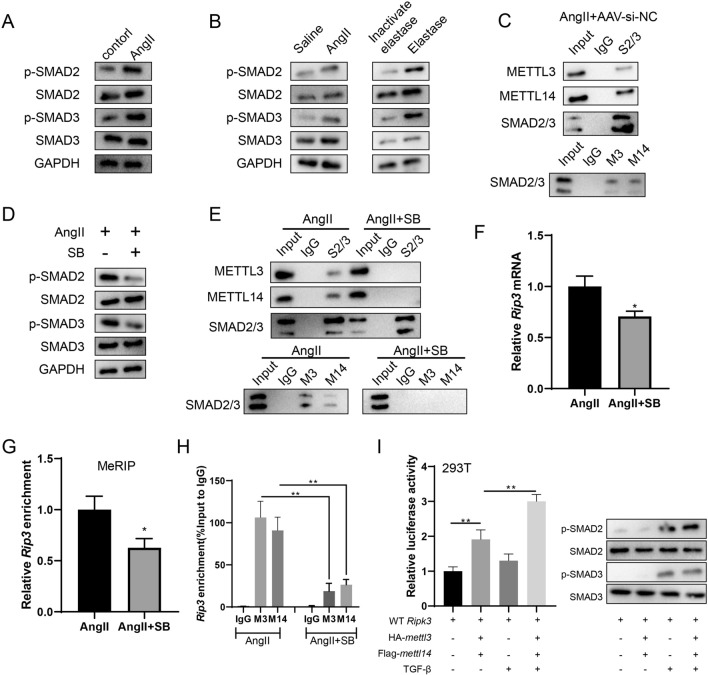
Fig. 6.
The activation of SMAD2/3 promotes the METTL3/METTL14-mediated m6A modification of rip3 mRNA. A VSMCs were divided into the Control group and the Ang II group. In the Ang II group, VSMCs were incubated with 200 nM Ang II. The protein expressions of SMAD2, p-SMAD2, SMAD3, and p-SMAD3 were determined by western blotting. B The protein expressions of SMAD2, p-SMAD2, SMAD3, and p-SMAD3 in mouse aortic wall tissues were determined by western blotting. C The binding between SMAD2/3 and METTL3/METTL14 in aortic wall tissues from the Ang II + AAV-si-NC group was confirmed using the Co-IP assay. D In VSMCs, after the inhibition of SMAD2/3 phosphorylation using 10 μM SB431542 (SB), the protein expressions of SMAD2, p-SMAD2, SMAD3, p-SMAD3 were determined by western blotting. E In VSMCs, after the inhibition of SMAD2/3 phosphorylation using 10 μM SB, the binding between SMAD2/3 and METTL3/METTL14 was determined using the Co-IP assay. F In VSMCs, after the inhibition of SMAD2/3 phosphorylation using 10 μM SB, the mRNA expression of rip3 was detected using the qRT-PCR. gapdh was used as the internal control. Relative gene expression was calculated using the 2−△△Ct method (n = 3, unpaired two-tailed t-test, *P < 0.05 vs Ang II). G In VSMCs, after the inhibition of SMAD2/3 phosphorylation using 10 μM SB, the m6A-bound rip3 enrichment in 3’UTR was detected using MeRIP (n = 3, unpaired two-tailed t-test, *P < 0.05 vs Ang II). H In VSMCs, after the inhibition of SMAD2/3 phosphorylation using 10 μM SB, the binding between rip3 mRNA with METTL3/METTL14 was detected using the RIP assay (n = 3, one-way ANOVA with Tukey’s post-hoc test, **P < 0.01). I In 293 T cells, after the activation of SMAD2/3 using 100 pM TGF-β1, the binding between rip3 mRNA with METTL3/METTL14 was detected using the dual-luciferase reporter assay (n = 3, one-way ANOVA with Tukey’s post-hoc test, **P < 0.01)

