Figure 5.
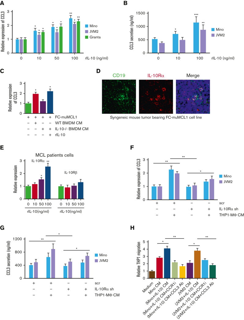
IL-10 regulates tumor-CCL3 expression. (A) Mino, JVM2, and Granta were treated with indicated concentrations of rIL-10 for 48 hours, and CCL3 expression was assessed using qRT-PCR. (B) Mino and JVM2 cells were treated with indicated concentrations of rIL-10 for 48 hours, and secretion of CCL3 was assessed using ELISA. (C) FC-muMCL1 cells were treated with CM collected from WT or IL-10-/- BMDM treated with or without rIL-10, and the expression level of CCL3 was assessed using qRT-PCR. (D) Immunofluorescent staining was performed on the syngeneic mouse tumors bearing FC-muMCL1 (n = 3) using CD19 (green) and IL-10Rα (red) antibodies. Nuclei were stained with DAPI (blue). A representative experiment is shown. (E) Cells of patients with MCL (n = 5) were treated with indicated concentrations of rIL-10 for 48 hours, and the mRNA level of IL-10Rα and IL-10Rꞵ were assessed using qRT-PCR. (F-G) THP1-MΦ CM was used to treat Mino and JVM2 cells infected with IL-10Rα shRNA or scramble shRNA for 72 hours, and the CCL3 mRNA level (F) and CCL3 secretion (G) were assessed using qRT-PCR and ELISA, respectively. (H) THP1 cells were incubated with CM collected from Mino or JVM2 pretreated with or without 100 ng/mL rIL-10 for 48 hours with 1μM CCR1 inhibitor or 0.5 μg/mL anti-CCL3 antibody, and the migration was assessed using chemotaxis assay. Data presented are representative of 3 independent experiments unless stated otherwise (∗P < .05; ∗∗P < .01).

