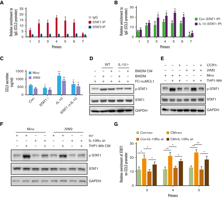
Figure 6.
Molecular mechanism of CCL3 regulation by IL-10. (A) ChIP assay was performed to detect enrichment of STAT1 and STAT3 on CCL3 promoters in Mino cells as described in the method section. (B) Mino cells were treated with or without 100 ng/mL rIL-10 for 24 hours, and the enrichment of STAT1 on the CCL3 promoter was detected using ChIP assay. (C) Mino and JVM2 cells were treated with 100ng/ml rIL-10 and/or 1μM STAT1 inhibitor fludarabine for 48h and the secretion of CCL3 was assessed using ELISA. The data presented are representative of 3 independent experiments. ∗P < .05; ∗∗P < .001. (D) FC-muMCL1 cells were cocultured with BMDM from WT or IL-10-/- mice (n = 3) or treated with CM collected from WT and IL-10-/- BMDM (n = 3), and the protein level of phospho-STAT1 and STAT1 were assessed using western blotting. (E) Mino or JVM2 cells were cocultured with THP1-MΦ with or without 1μM CCR1 inhibitor BX-471 for 24h, and the protein level of phospho-STAT1 and total STAT1 were assessed using western blotting. The experiment was repeated 3 times with similar results and representative data shown. (F) Mino and JVM2 cells infected with IL-10Rα shRNA or scramble shRNA for 72 hours were incubated with THP1-MΦ CM, and the protein level of phospho-STAT1 and total STAT1 was assessed using western blotting. (G) Mino cells infected with IL-10Rα shRNA or scramble shRNA for 72 hours were incubated with THP1-MΦ CM, and the enrichment of STAT1 on the CCL3 promoter was assessed using ChIP assay. ∗P < .05; ∗∗P < .01. Data presented are representative of 3 independent experiments unless stated otherwise (∗P < .05; ∗∗P < .01; ∗∗∗P < .001).

