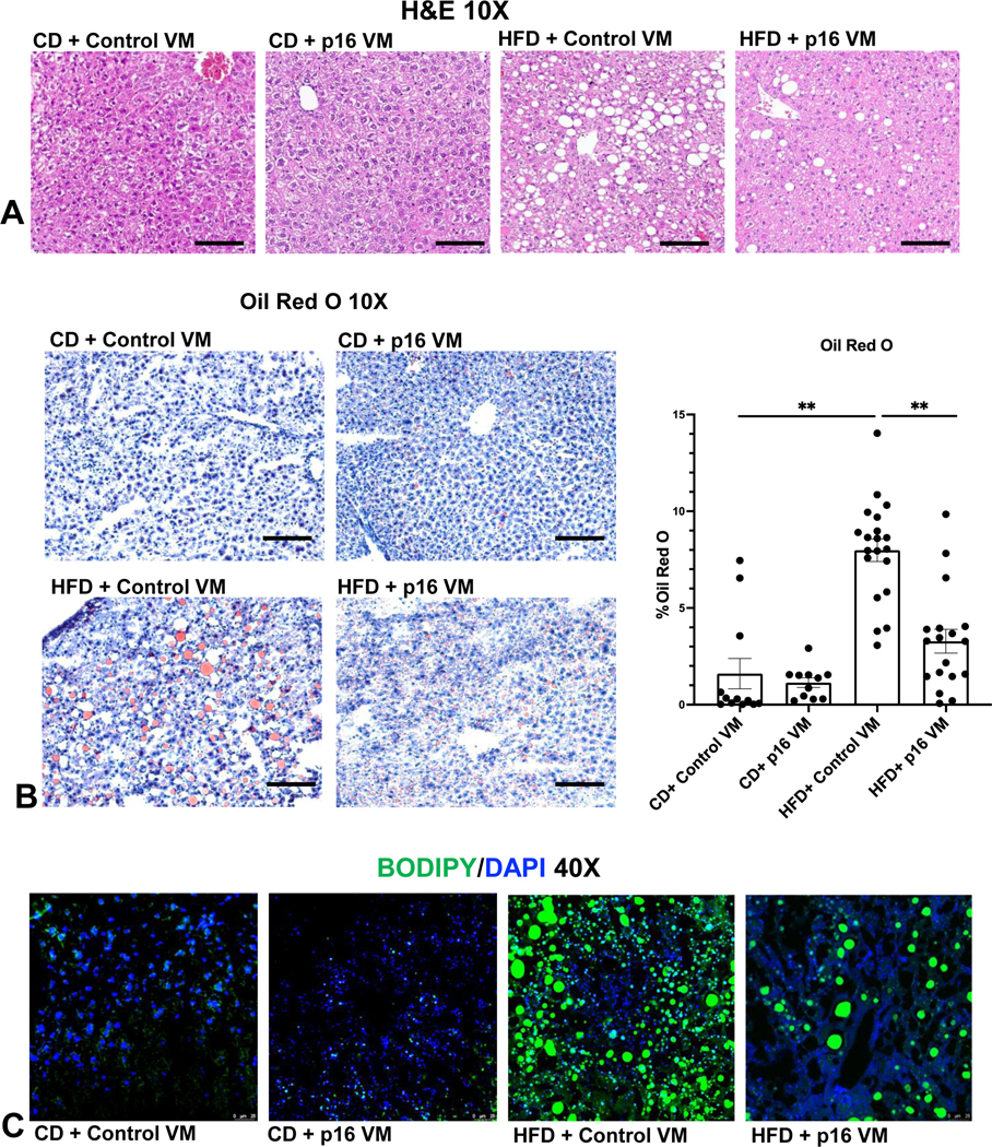
Figure 2: p16 VM reduced steatosis in WT HFD fed mice.
By H&E staining we found that WT HFD mice had moderate steatosis with stage 1–2 portal fibrosis, ballooning hepatocytes, inflammation, and ductular proliferation compared to CD fed mice treated with control VM. Treatment with p16 VM in WT HFD fed mice displayed patchy ductular proliferation and steatosis, the absence of ballooning hepatocytes and minimal stage 1–2 fibrosis as shown by H&E (10x); there were no noticeable differences between the CD control VM and CD p16 VM mice (A). Oil Red O staining (10x) showed a significant increase in % lipid droplet area (steatosis) in HFD control VM mice compared to CD control VM mice and WT HFD mice treated with p16 VM had a significant reduction in lipid droplet presence (B). BODIPY staining (40x) demonstrated increased neutral lipid droplet presence in WT HFD control VM mice compared to CD control VM which decreased in HFD p16 VM mice (C). Data are expressed as mean ± SEM. Each dot represents an image; n = 12–20 images from n = 4–6 mice for Oil Red O were used for semi-quantification. Representative images for stains were chosen from n = 12–20 images per group for (B). **P<0.05.

