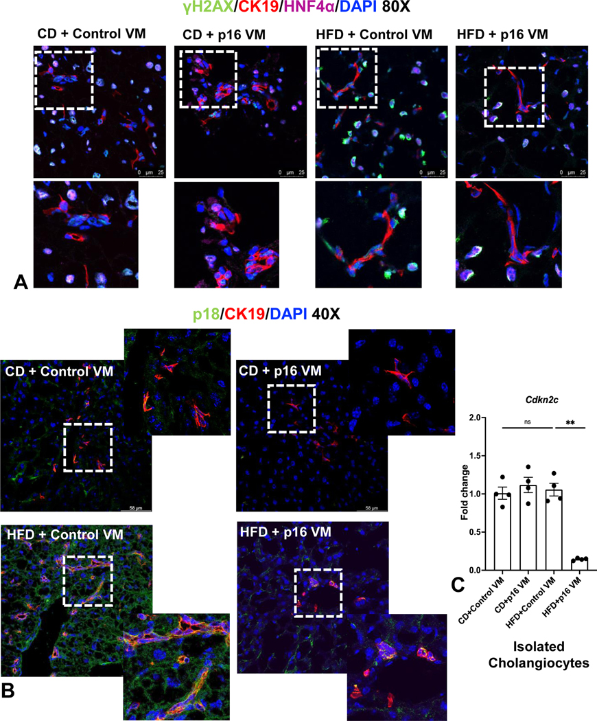
Figure 4: p16 VM reduced hepatocyte and cholangiocyte senescence in WT HFD fed mice.
γΗ2Α.X (green) staining was elevated in both hepatocytes (cyan) and cholangiocytes (red) of HFD control VM mice and HFD p16 VM mice had reduced γΗ2Α.X staining in both cholangiocytes and hepatocytes (A). p18 immunostaining (co-stained with CK-19) increased in HFD control VM mice compared to CD control VM and when p16 VM was administered to HFD mice, p18 expression decreased (B). By qPCR, gene expression of p18 (Cdkn2c) did not change between WT CD control or p16 VM or WT HFD control VM; however, there was significant reduction of p18 expression in WT HFD mice treated with p16 VM (C). Data are expressed as mean ± SEM. Each dot of qPCR represents a technical replicate of 4 reactions from n = 4–6 mice and normalized with Rps18 as the housekeeping gene. Immunostaining of γΗ2Α.X (80x) and p18 (40x) are representative of at least 4 different sections of tissues per treatment group. **P< 0.001; ns = non-significant.

