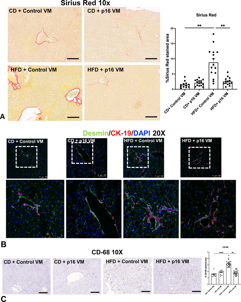
Figure 5: p16 VM treatment reduced collagen deposition, HSC activation and inflammation in WT HFD mice.
WT HFD control VM mice displayed a significant increase in Sirius Red staining/collagen deposition compared to WT CD control VM mice, which was significantly reduced in WT HFD p16 VM mice (A). Co-immunostaining of desmin (green) with CK-19 (red) indicated increased immunoreactivity of desmin (20x) in the portal area in WT HFD control VM mice compared to CD control VM mice, whereas p16 VM treatment decreased desmin immunoreactivity in WT HFD fed mice (B). CD-68 positivity significantly increased in WT HFD control VM mice compared to CD control VM mice and CD-68 positive cells significantly decreased in WT HFD p16 VM mice (C). Data are expressed as mean ± SEM. Each dot represents an image; n = 12–15 images from n = 4–6 mice were used for Sirius Red (10x) and n = 10–20 images for CD-68 (10x) semi-quantification. Representative images of desmin/CK-19 staining were obtained from at least n = 6 images per group. ****P< 0.001; ** P< 0.05.

