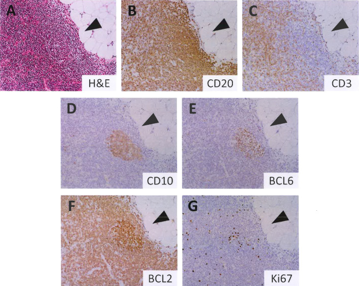
Fig. 1.
Histopathology of isolated in situ follicular B-cell neoplasm (ISFN). Hematoxylin and eosin staining showing a lymphoid follicle with a monotonous germinal center (GC) (A). GC is involved by ISFN, in which neoplastic B cells are CD20+ (B), CD3- (C), CD10+ (D), BCL6+ (E), and BCL2+ (F). The ISFN lesion exhibits more intense BCL2 staining than the background B and T cells. The Ki67-labeling index is low (G).

