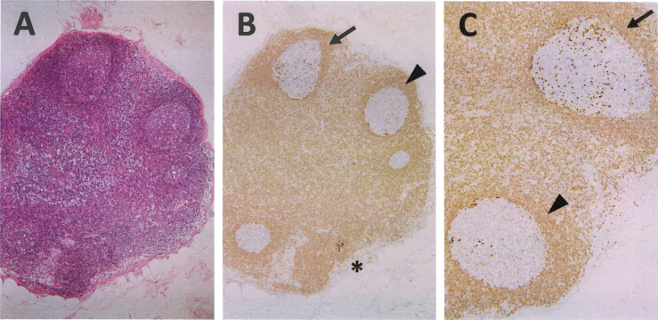
Fig. 3.
GCs variably involved by ISFN. (A) Hematoxylin and eosin staining demonstrates reactive-looking GCs. (B and C) Immunohistochemistry for BCL2 highlights a clear ISFN lesion (*) and GCs with scattered ISFN cells strongly expressing BCL2 (arrow and arrowhead). Note that the number of BCL2-positive cells in GCs are variable as indicated by the arrow and arrowhead.

