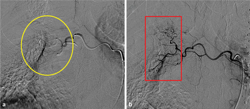
Fig. 5.

Recanalization in bronchial artery embolization with microspheres. ( a ) Immediate postprocedural angiogram demonstrates relatively proximal vessel occlusion (yellow circle). Distal irregular vasculature is no longer identified. ( b ) Repeat bronchial angiogram a few days later demonstrates recanalization (red box) and recurrent hemorrhage, likely secondary to proximal particle clumping on the initial intervention leading to a false endpoint.
