Fig. 6.

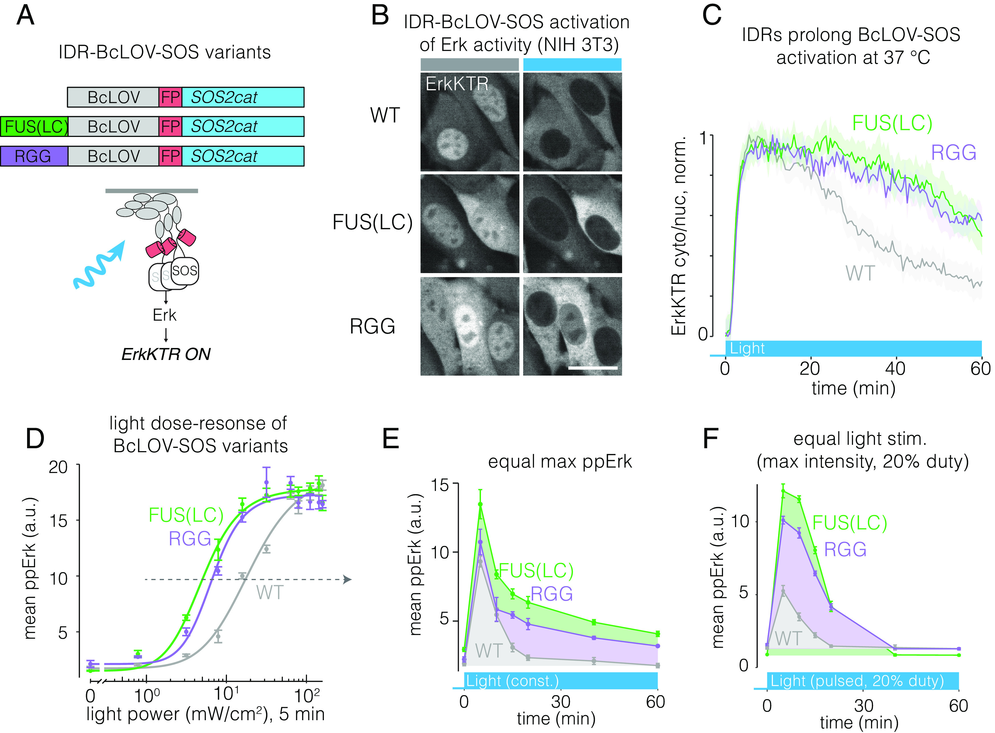
IDRs enhance sensitivity and strength of BcLOV-SOScat. (A) IDR-fused variants of BcLOV-SOScat. (B) The Ras/Erk pathway was activated in cells by IDR-BcLOV-SOScat variants in NIH 3T3 fibroblasts, as measured by the ErkKTR reporter. (Scale bar, 20 μm.) (C) Quantification of ErkKTR activity during 1 h of stimulation by BcLOV-SOScat variants. IDR variants show slower pathway decay. See SI Appendix, Table S1 for details of optogenetic illumination parameters. (D) Dose–response of light intensity on ppErk after 5 min of constant illumination at the indicated light dose. Data represent mean ± SEM of four replicates, each representing the mean signal from ~200 to 1,000 cells. (E) Comparison of ppErk activation dynamics by BcLOV-SOScat variants, each stimulated at a constant light intensity that produced equivalent max ppErk, as determined in (D) (dotted arrow). IDR variants showed higher sustained and integrated signaling over 1 h of stimulation. Data represent the mean ± SEM of four replicates, each representing the mean of ~50 to 400 single cells. (F) Comparison of ppErk activation dynamics by BcLOV-SOScat variants in response to pulsatile (20% duty cycle) maximum intensity light. IDR variants achieved >twofold higher maximal signal and more sustained and integrated activity compared to wt BcLOV-SOScat. Data represent mean ± SEM of four replicates, each representing the mean of ~100 to 600 cells.
