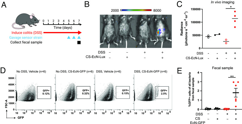
Fig. 3.
Calprotectin-sensing EcN can reliably detect gut mucosal inflammation in vivo in the DSS-induced colitis mouse model. (A) Mice were gavaged daily with 3% DSS for 7 d to induce colitis. CS EcN-Lux or CS EcN-GFP was gavaged daily for 3 d prior to IVIS imaging or fecal sample collection. (B) Representative live animal luminescence imaging was performed using IVIS Spectrum Instrument on mice (n = 4) that were gavaged CS EcN-Lux compared to controls that were not treated with DSS or not gavaged CS EcN-Lux (n = 2). (C) Luminescence was quantified for live animal imaging of mice gavaged with CS EcN-Lux with colitis induced by DSS (n = 4) and controls (n = 2). (D) Representative flow cytometry images quantifying GFP positive cell populations in mice that were treated with DSS and gavaged CS EcN-GFP and controls. Carbenicillin was given 2 d prior to bacterial gavage. (E) GFP expression was quantified by flow cytometry from stool of mice gavaged with CS EcN-GFP with colitis induced by DSS (n = 6) and controls (n = 6). **P < 0.01 and ***P < 0.001 for Student’s unpaired t test for indicated comparisons. Data are represented as mean ± SEM.

