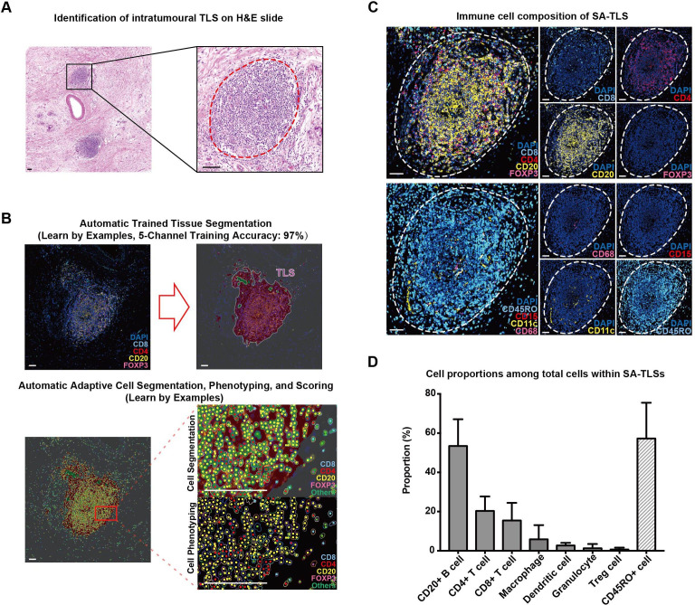
Figure 1.
Representative structure of intratumoral tertiary lymphoid structures (TLSs) in surgery alone (SA) patients. (A) H&E staining images of intratumoral TLSs in pancreatic ductal adenocarcinoma tumor tissues; TLS is circled with dotted red line; scale bar: 100 µm. (B) The segmentation of different cell types within TLSs using inForm software; scale bar: 50 µm. (C) Fluorescent multiplex immunohistochemistry staining images combining CD4 (for CD4+ T cells), CD8 (for CD8+ T cells), CD20 (for CD20+ B cells) and FOXP3 (for Treg cells) in one tissue section and CD68 (for macrophages), CD15 (for granulocytes), CD11c (for dendritic cells) and CD45RO in another serial section; TLSs are circled with dotted white lines; scale bar: 50 µm. (D) The proportions of seven immune cells and CD45RO+ cells within intratumoral TLSs in the 80 TLS (+) samples in the SA group.

