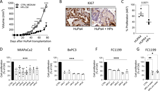
Fig. 2.
Effect of Ps conditioned media on PDAC growth. (A) Growth curve of HuPa4 tumors transplanted s.c and treated with HPs conditioned medium or control medium (mean ± SEM; n = 4 for each group, two-way ANOVA with Bonferroni’s multiple comparison test, *p < 0.005,**p < 0.0005, ***p < 0.0001). Ki67 immunostaining (B) and quantification of tumor cell proliferation (C) at euthanasia (400X, scale bar 100 μm) (mean ± SEM). Proliferation of MIAPaCa2 (D), BxPC3 (E), and FC1199 cells (F) in response to respectively 7 and 5 human and murine Ps CM, (72 h of treatment). Data are percentages of control from at least two different experiments (mean ± SEM; one-way ANOVA with Dunnett’s multiple comparison test, ***p < 0.001 ****p < 0.0001). (G) Proliferation of FC1199 cells in response to murine Ps CM, containing or not Langerhans islets. Data are percentages of control from at least five different Ps preparations (mean ± SEM; one-way ANOVA with Dunnett’s multiple comparison test, **p < 0.01, *p < 0.05)

