Fig. 4.
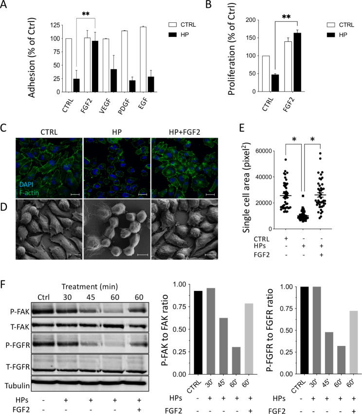
FGF2 blocks the de-adhesive effect of HPs CM on MIAPaCa2 cells. MIAPaCa2 cells were treated with HPs CM together with the growth factors (20 ng/mL). (A) Quantification of cell adhesion after 1 h of treatment (mean ± SEM; one-way ANOVA with Dunnett’s multiple comparison test, **p < 0.05). (B) Quantification of cell proliferation after 72 h of treatment (mean ± SEM; one-way ANOVA with Dunnett’s multiple comparison test, **p < 0.005). (C) Immunofluorescence analysis of F-actin (green, scale bar 25 μm) and (D) Scanning electron microscopy (SEM) (scale bar 10 μm) after 1 h of treatment with HPs CM with or without FGF (20 ng/mL). (E) Analysis of single cell area after 1 h of treatment (mean ± SEM; one-way ANOVA with Dunnett’s multiple comparison test, *p < 0.001). (F) Western Blot analysis of P-FAK, T-FAK, P-FGFR, T-FGFR and tubulin in MIAPaCa2 cell lysates after treatments and quantification of FAK and FGFR phosphorylation expressed as the ratio of phosphorylated protein to total protein

