Abstract
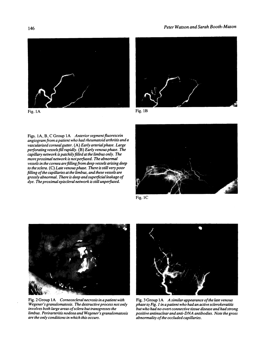
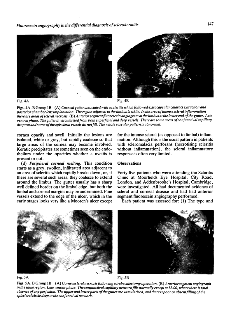
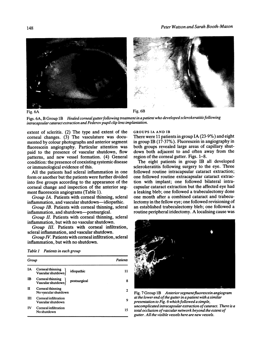
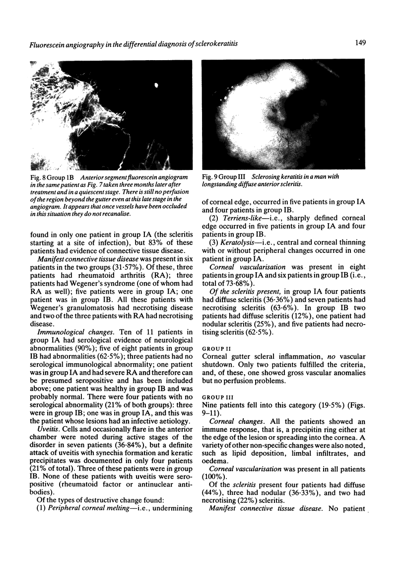
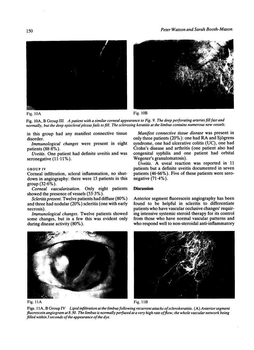
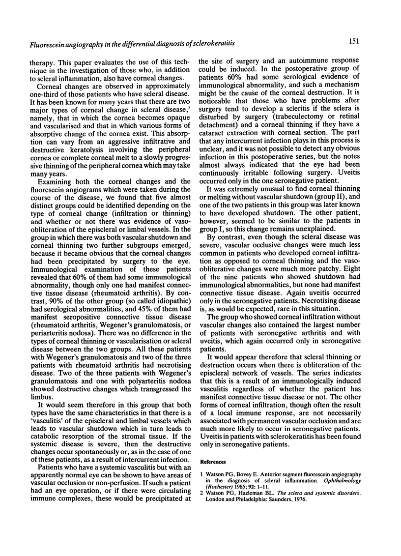
Anterior segment fluorescein angiography has been used in the investigation of patients with sclerokeratitis. This showed that corneal thinning or destruction was associated with non-perfusion of the episcleral vasculature. The changes arose either as a result of a systemic vasculitis in seropositive individuals or were induced by surgery to the eye. Infiltrative forms of sclerokeratitis were commoner in seronegative patients and were less often associated with vascular shutdown.
Full text
PDF
Images in this article
Selected References
These references are in PubMed. This may not be the complete list of references from this article.
- Watson P. G., Bovey E. Anterior segment fluorescein angiography in the diagnosis of scleral inflammation. Ophthalmology. 1985 Jan;92(1):1–11. doi: 10.1016/s0161-6420(85)34074-5. [DOI] [PubMed] [Google Scholar]


