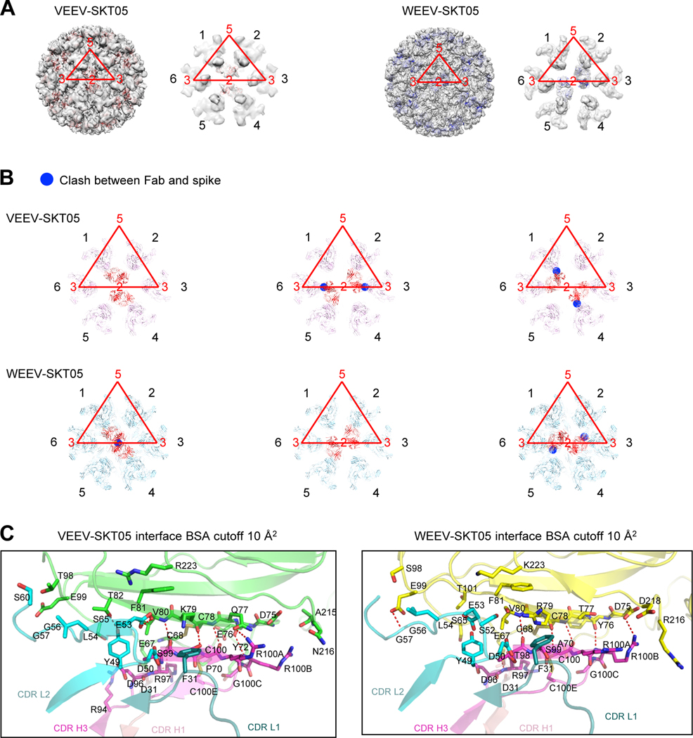
Figure 5. SKT05 utilizes different symmetry to bind VEEV and WEEV.
(A) Overall view of the reconstruction density for VEEV and WEEV VLPs with bound SKT05 Fab and close-up view of icosahedral 2-fold axis surrounded with six E1:E2 spikes (labeled 1–6 in black font, with VLP symmetry axes labeled in red font, and the icosahedral asymmetric unit also shown in red). SKT05 Fabs are shown as red and blue ribbons, respectively. SKT05 bound to spikes labeled 1 and 4 in VEEV and to spikes labeled 2 and 5 in WEEV. (B) VEEV-SKT05 and WEEV-SKT05 complexes docked into spike reconstruction density surrounding 2-fold axis. Clashes are highlighted with blue circles and were observed between SKT05 and nearest polypeptide chains when VEEV-SKT05 was docked to spike pairs 2,5 and 6,3, and when WEEV-SKT05 was docked to spike pairs 1,4 and 6,3. (C) Details of interactions between VEEV and WEEV VLPs with SKT05 are shown in ribbons, with all interactions with a BSA larger than 10 Å2 shown in sticks. Left panel: interactions of VEEV E1 (green) with CDR-H1, CDR-H3, CDR-L1 and CDR-L2 of SKT05. Right panel: interactions of WEEV E1 (yellow) with CDR-H1, CDR-H3, CDR-L1 and CDR-L2 of SKT05. See also Table S3 and Data S1.

