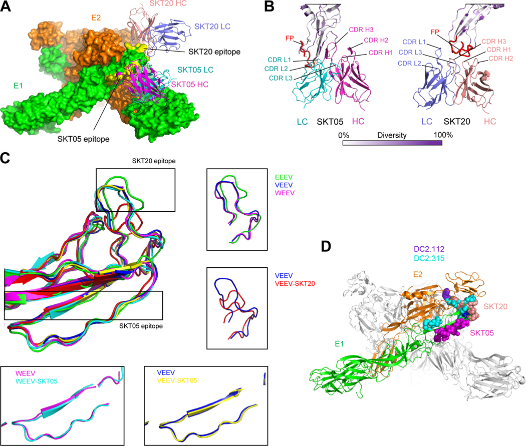
Figure 6. Structural details of SKT05 and SKT20 broad recognition.
(A) Overall view of the WEEV spike bound by SKT05 and SKT20. Molecular surfaces are shown for the E1:E2 trimer (colored green and orange, respectively), with SKT05 and SKT20 variable domains displayed in ribbons and their epitopes highlighted in yellow. (B) Close-up view of SKT20 and SKT05 in contact with WEEV E1 glycoprotein drawn in ribbons and colored by sequence diversity according to the white-to-purple key, with two conformations of the fusion peptide highlighted in red. (C) Superposition of E1 glycoproteins for EEEV, VEEV, WEEV, and their SKT05 and SKT20-bound complexes. In free EEEV, VEEV, and WEEV VLPs, E1 glycoproteins have a similar conformation for the fusion peptides. Binding of SKT05 does not affect the conformation of E1, whereas binding of SKT20 results in a dramatic change in the conformation of the fusion peptide. (D) View of the WEEV spike, displayed in ribbons representation, with one E1:E2 protomer colored green and orange, respectively, and the two other protomers colored medium and light gray. Epitopes for SKT05, SKT20, DC2.112 and DC2.315 are shown in sphere representation, and colored magenta, orange, purple, and cyan, respectively. See also Data S1.

