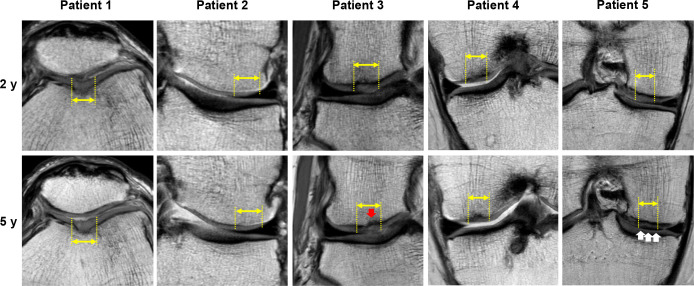
Figure 3.
Proton density-weighted MRI scans of the injured cartilage sites (yellow double arrows and dotted lines) at 2 and 5 years postoperatively for all study patients. The red arrow in patient 3 indicates a bony defect of the subchondral bone area. The white arrows in patient 5 indicate the abnormal signal intensity of the repaired cartilage. MRI, magnetic resonance imaging.

