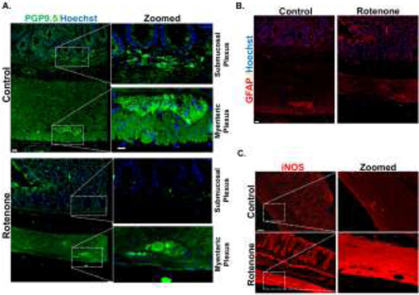
Figure 6: Enteric neuronal degeneration in the submucosal plexus in a rat rotenone (Rot) model of PD.
(A) IHC of rat colon treated with Rot (2.8mg/kg/i.p) showing decreased expression of PGP 9.5 in the submucosal plexus. (B) IHC of rat colon treated with Rot showing no significant change in expression of GFAP in the myenteric plexus. (C) IHC of rat colon treated with Rot showing increased expression of iNOS in the myenteric plexus (n=2). Scale bar, 20 μm.

