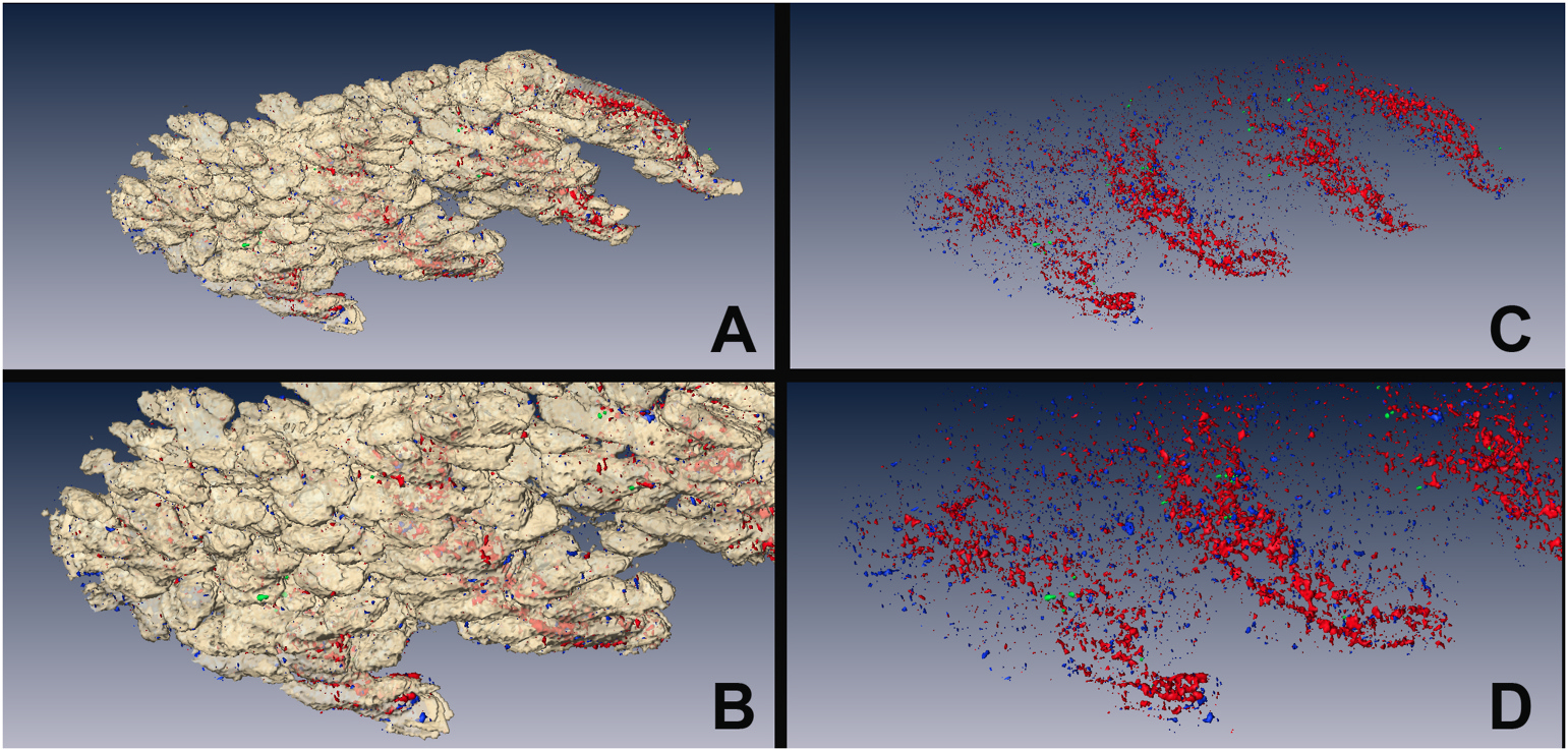
Fig. 5.

Using the segmented region of the meibomian gland as a mask, the non-meibomian gland eyelid tissue was extracted from the 3D reconstruction to reveal the shape of the gland (A and B, white shell) and the spatial distribution of GFP-label-retaining cells (Green), Ki67-proliferating cells (Blue), and E-cadherin-positive ductal epithelium (Red) as shown in C and D.
