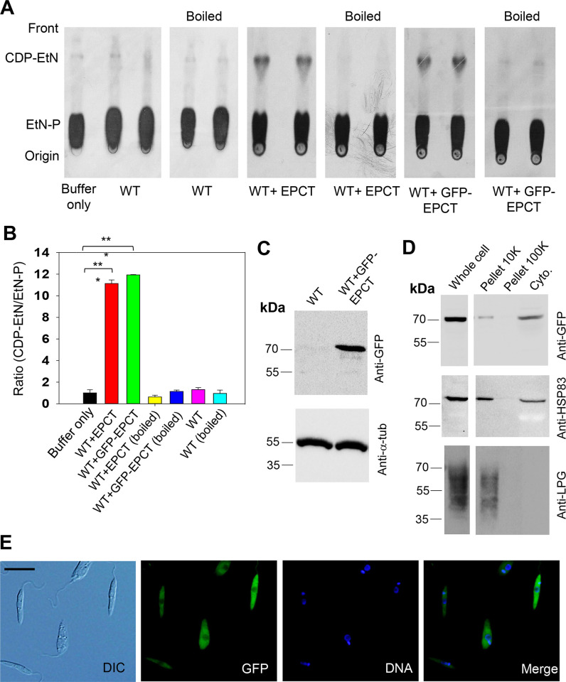
Fig 2. L. major EPCT is a functional enzyme located in the cytoplasm.
(A-B) Whole cell lysates (boiled and not boiled, two repeats were shown) of WT, WT+EPCT, and WT+GFP-EPCT promastigotes were incubated with [14C]-EtN-P followed by TLC analysis as described in Materials and Methods. Representative TLC images were shown in A and the average ratios of [14C]-CDP-EtN]:[14C]-EtN-P were normalized to WT (as 1.0) and summarized in B (error bars represent standard deviations from four repeats, ***: p < 0.001). (C) Promastigote lysates of WT and WT + GFP-EPCT were analyzed by western-blot using an anti-GFP antibody (top) or anti-α-tubulin antibody (bottom)(D) Whole cell lysate (1.0 x 10⁶ cells) and sub-cellular fractions of WT + GFP-EPCT cells were probed by anti-GFP (top), -HSP83 (middle), or -LPG (bottom) antibodies. (E) Log phase promastigotes of WT+GFP-EPCT were examined by fluorescence microscopy. DIC: differential interference contrast. Scale bar: 10 μm. DNA: staining with Hoechst 33342. Merge: overlay of GFP and DNA.

