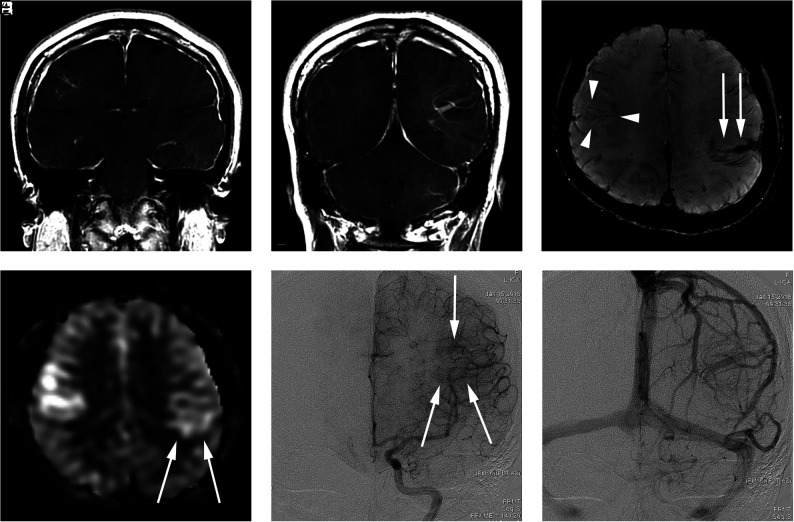
FIG 2.
A 34-year-old woman presenting with a headache (case 6). A and B, Contrast-enhanced T1-weighted coronal MR imaging shows a DVA-like lesion in the bilateral parietal lobes. C, SWI demonstrates only hypointense signal in both the right (arrowheads) and left (arrow) lesions. D, The ASL quantitative CBF image demonstrates mildly hyperintense signal intensity in the parenchyma, corresponding to the location of the lesion. The left-sided lesion exhibits particularly subtle signal (arrows). E, In the late arterial phase of DSA, dilated medullary veins are gradually and subtly visualized (arrows). F, In the venous phase, dilated medullary veins draining to a collecting vein typical of a DVA are seen.

